- Published on
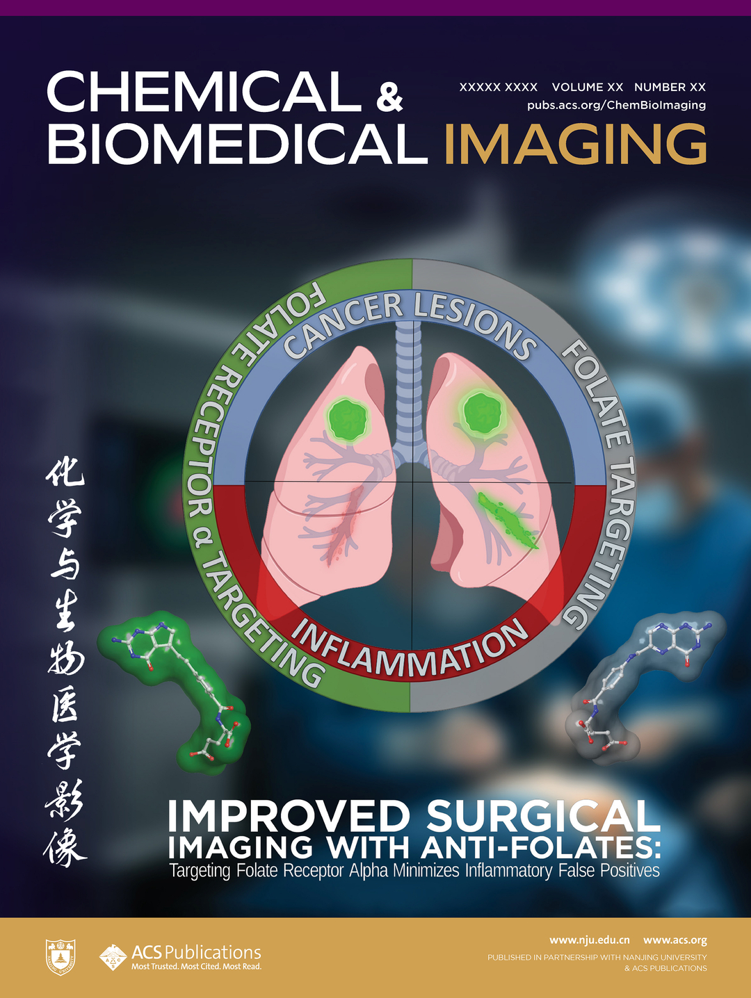
Chemical & Biomedical Imaging - ML-Optimized Diagnostic Probe for Tumor-Inflammation Discrimination

We've published work in Chemical & Biomedical Imaging covering our development of pemefolacianine (NY-07) to address a critical failure mode in fluorescence-guided surgery. Current FDA-approved probes generate false-positive rates of 25-68% because they accumulate in both tumor and inflammatory tissue, leading to unnecessary resections and surgical complications. NY-07, designed through ML optimization integrated with CADD modeling of antifolate drugs, achieved 8-fold selectivity for tumor over inflammatory tissue. The compound is currently being advanced through Phase II clinical trials by our industrial collaborator Nanjing Nuoyuan Medical Devices following IND approval from both FDA and NMPA.
The problem is mechanistic. Folate receptor α (FRα) is upregulated in approximately 40% of cancers, while folate receptor β (FRβ) is expressed on activated inflammatory macrophages. Existing folate-conjugated probes (including FDA-approved Pafolacianine - OTL-38) bind both receptors with comparable affinity, producing false signals in inflamed tissue adjacent to tumors. Surgeons compensate through visual inspection and palpation, adopting conservative resection strategies that increase surgical morbidity while still missing residual disease.
Our team approached this as a receptor selectivity optimization problem. Regression models trained on ChEMBL inhibitor datasets for FRα (CHEMBL2121) and FRβ (CHEMBL5064) screened a library of modified antifolate scaffolds. The models predicted IC50 values for both receptor subtypes, calculating selectivity ratios (FRβ IC50/FRα IC50) for each candidate. NY-07, derived from the pemetrexed scaffold with systematic modifications including tyrosine substitution, showed favorable predicted selectivity confirmed through molecular docking against the FRα crystal structure (PDB: 4LRH).
Experimental validation demonstrated Kd of 61.67 nM for FRα versus 486.9 nM for FRβ. In mouse xenograft models, NY-07 achieved tumor-to-background ratio of 3.23 ± 0.28 at 1 hour post-administration, maintaining clinically relevant imaging windows (TBR >1.5) for up to 12 days. The probe detected lesions smaller than 1 mm across multiple tumor types. Critically, in inflammation models (peritonitis, pneumonia, arthritis), tumor fluorescence intensity (3.57 ± 0.76 × 10^8 photons/sec/cm²/sr) significantly exceeded inflamed tissue signal (1.35 ± 0.16, p < 0.01). Immunohistochemistry confirmed FRα-positive tumor areas showed strong fluorescence while FRβ-positive inflammatory regions produced minimal signal.
The design-to-clinic timeline was under three years, demonstrating the power of ML-guided receptor selectivity optimization to rapidly advance clinically viable candidates.