Fluorescence Image Guided Surgery
Improving Surgical Outcomes Through Advanced Imaging Technologies
Our group pioneers cutting-edge image-guided surgery technologies that enable surgeons to visualize cancer with unprecedented clarity during operations. Through innovative algorithms and novel imaging approaches, we're transforming how surgeons detect, locate, and remove tumors while preserving healthy tissue.
Our group pioneers cutting-edge image-guided surgery technologies that enable surgeons to visualize cancer with unprecedented clarity during operations. Through innovative algorithms and novel imaging approaches, we're transforming how surgeons detect, locate, and remove tumors while preserving healthy tissue.
The Critical Need for Precision
Cancer surgery represents one of medicine's most significant challenges: surgeons must remove every cancerous cell while preserving as much healthy tissue as possible, often making difficult decisions that determine patient survival and quality of life. Traditional surgery relies heavily on the surgeon's experience and tactile feedback to distinguish between healthy and cancerous tissue, leading to incomplete tumor removal in some cases or excessive healthy tissue removal in others. Without real-time molecular-level guidance, surgeons face an inherent trade-off between thoroughness and preservation, resulting in variable outcomes that depend on individual expertise rather than standardized, data-driven precision. Our image-guided surgery research addresses this fundamental limitation by providing surgeons with intelligent imaging systems that reveal cancer at the molecular level during operations, enabling complete tumor removal while minimizing damage to healthy tissue and improving consistency across all surgical procedures.
Cancer surgery represents one of medicine's most significant challenges: surgeons must remove every cancerous cell while preserving as much healthy tissue as possible, often making difficult decisions that determine patient survival and quality of life. Traditional surgery relies heavily on the surgeon's experience and tactile feedback to distinguish between healthy and cancerous tissue, leading to incomplete tumor removal in some cases or excessive healthy tissue removal in others. Without real-time molecular-level guidance, surgeons face an inherent trade-off between thoroughness and preservation, resulting in variable outcomes that depend on individual expertise rather than standardized, data-driven precision. Our image-guided surgery research addresses this fundamental limitation by providing surgeons with intelligent imaging systems that reveal cancer at the molecular level during operations, enabling complete tumor removal while minimizing damage to healthy tissue and improving consistency across all surgical procedures.
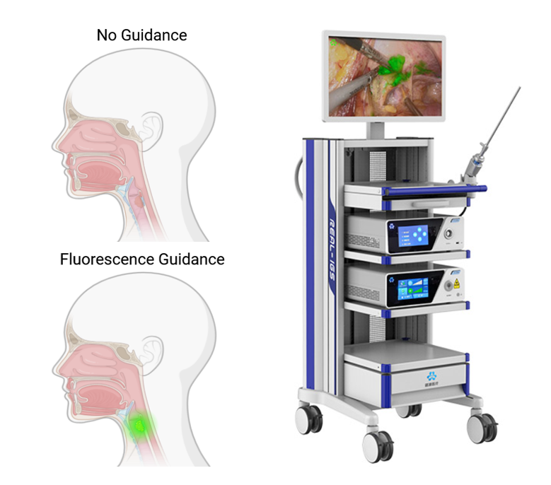
How Image-Guided Surgery Works: Image-guided surgery operates through three integrated components during operations. The camera system uses near-infrared sensors that detect fluorescent signals outside the visible spectrum, mounted on surgical microscopes or handheld devices that display video feeds to operating room monitors. The fluorescent dye is administered to patients before surgery—either as contrast agents that accumulate in tumors through vascular permeability differences, or as targeted probes that bind to specific cancer cell receptors like EGFR—producing fluorescent signals that differentiate cancerous from normal tissue. Data processing algorithms enhance these fluorescent signals in real-time, improving signal-to-noise ratios, defining tissue boundaries, and overlaying fluorescent information onto standard surgical video, providing surgeons with additional visual information about tissue characteristics during tumor removal procedures.
Our Research Areas:Advanced Algorithm Development Signal processing and machine learning techniques for real-time tumor boundary detection and surgical image enhancement.
Novel Fluorescent Targeting Systems Near-infrared probes that bind to tumor receptors for tissue localization and lymph node mapping.
Clinical Translation Moving laboratory research to operating room practice through clinical trials and regulatory development.
Novel Fluorescent Targeting Systems Near-infrared probes that bind to tumor receptors for tissue localization and lymph node mapping.
Clinical Translation Moving laboratory research to operating room practice through clinical trials and regulatory development.
Featured Publications:
"Fluorescence Discrimination of Cancer from Inflammation by Selective Targeting Folate Receptor α"
Chemical & Biomedical Imaging (2025)
Details our development of Pemefolacianine, now in Phase II trials for selective cancer imaging.
"Signal-to-Noise Ratio Imaging and Real-Time Sharpening of Tumor Boundaries for Image-Guided Cancer Surgery"
Analytical Chemistry (2025)
Signal processing techniques that improve tumor boundary visualization during surgery.
DOI: 10.1021/acs.analchem.5c00530
"A Novel Near-Infrared EGFR Targeting Probe for Metastatic Lymph Node Imaging"
Journal of Nanobiotechnology (2023)
Cancer-specific imaging probe that binds to EGFR receptors, enabling detection of metastatic lymph nodes in preclinical models.